General Information
- Primary intraosseous epithelial neoplasm
- Low-grade malignancy and predilection for tibia
- Associated with intraosseous fibro-osseous proliferation (osteofibrous dysplasia)
- Classic vs. Differentiated
- Classic – grow beyond cortex, older patients, sometimes metastasize
- Differentiated – confined to cortex, earlier age, do not metastasize
Clinical Presentation
Signs/Symptoms:
- pain & Swelling, painless swelling, or pain alone
- 50% have history of localized trauma
Prevalence:
- males and females affected equally
Age:
- range 3-72 years old
- ~50% present in second or third decade
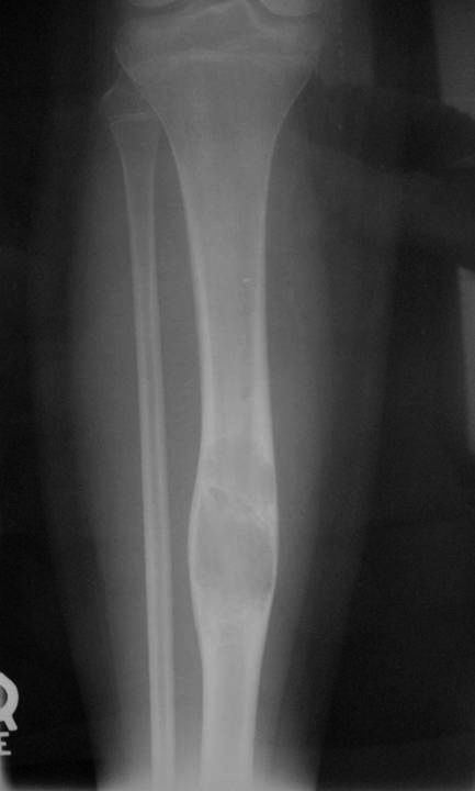
Sites:
- Predilection for tibia (90%)
- favoring diaphyseal portion of bone
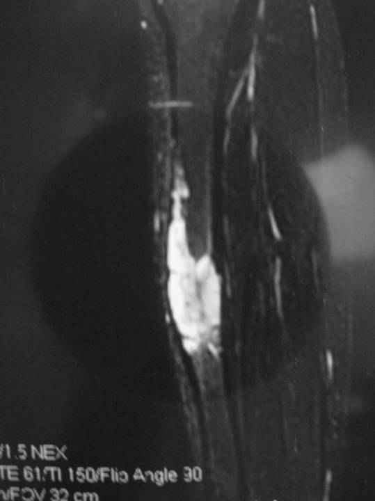
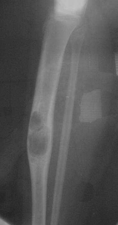
Radiographic Presentation
- Sharply defined osteolytic defect (lobulated, multicystic, or “soap bubble”)
- May be considerable perilesional sclerosis
Gross Pathology
- Sharp demarcation and lobulated configuration may be seen
- Solid areas are soft to firm, gray or white, granular or fibrous
- Cystic spaces and areas of intralesional hemorrhage are common
- Length: 5 cm to entire shaft
Microscopic Pathology
- Four histologic patterns:
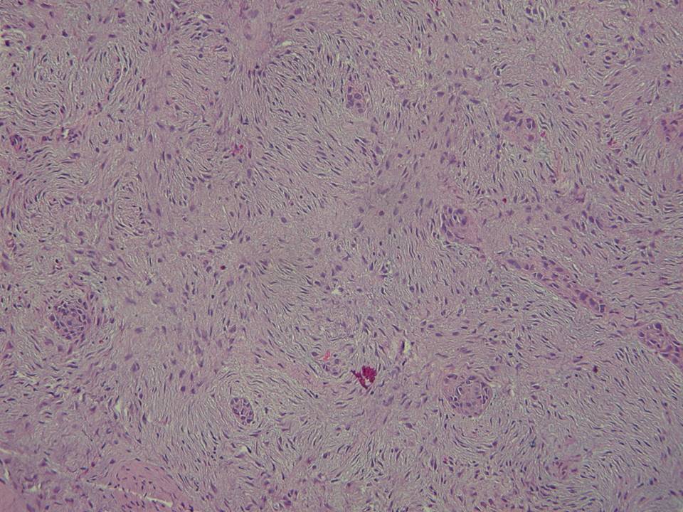
- spindled
- basaloid
- tubular
- squamoid
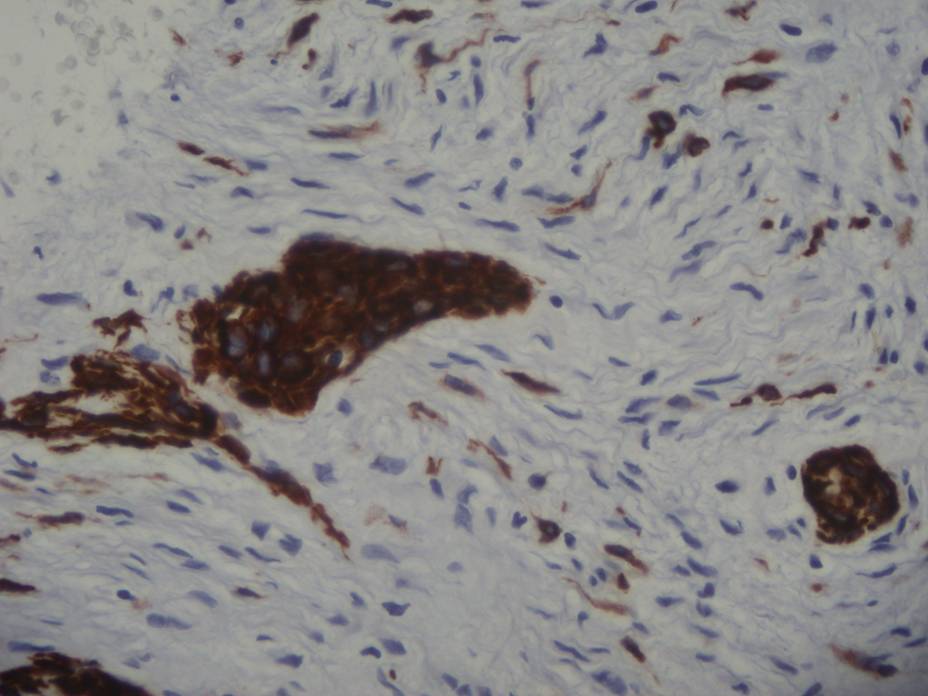
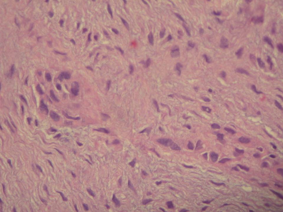
- Situated in a loose or dense fibrous stroma
- May form tubular structures lined by one or two cells, which may branch and anastomose
- Appear as small, vascular tubules with lumens in cross section
- May have squamous differentiation
- Nuclei are usually bland