GENERAL INFORMATION
Giant cell tumor of tendon sheath is defined as a benign reactive lesion which is similar to PVNS. Giant cell tumor of tendon sheath is also named tenosynovial giant cell tumor. GCT of tendon sheath is a circumscribed tumor that does not always arise from the tendon sheath but may arise from the synovium. Most common in patients after 30 years old and it is the second most common benign hand tumor after a ganglion cyst. GCTTS present as localized or diffuse proliferation of synovial-like cells, giant cells, inflammatory cells and xanthoma cells along tendon sheaths. Must be differentiated from diffuse tenosynovial giant cell tumor. Never metastasize but local recurrence after excision varies from 4% to 44%.
CLINICAL DATA
• It is a slowly growing and painless lesion of soft tissues.
• Second most common tumor of the hand
• Occurs commonly on the volar aspect of fingers and hand
• Affects individuals between 30 and 50 years old
Differential diagnosis
• Clear cell sarcoma
• Fibroma of tendon sheath
• Epithelial inclusion cyst
• Fibrous histiocytoma of skin (dermatofibroma)
• Epithelial sarcoma
CLINICAL PRESENTATION
Signs/Symptoms
• Slowly growing mass in the hand
• Usually asymptomatic
• Mass usually found in soft tissue near tendon
Prevalence
• Fairly common
• Slight predilection for women
Age
• 3rd-5th decades
Sites
• Hand and wrist m/c locations (65-89%)
• Foot and ankle (5-15%)
• Pressure erosions in 15% (ankles/feet)
• Can erode adjacent bone and destroy it
RADIOGRAPHIC PRESENTATION
Plain x-ray
• Soft tissue mass
• Pressure erosions in underlying bone in 15% of cases
• Calcifications are uncommon
MRI
• Isointense to muscle T1
• Low to intermediate T2
• May bloom on gradient echo (hemosiderin deposition)
• May demonstrate intense enhancement
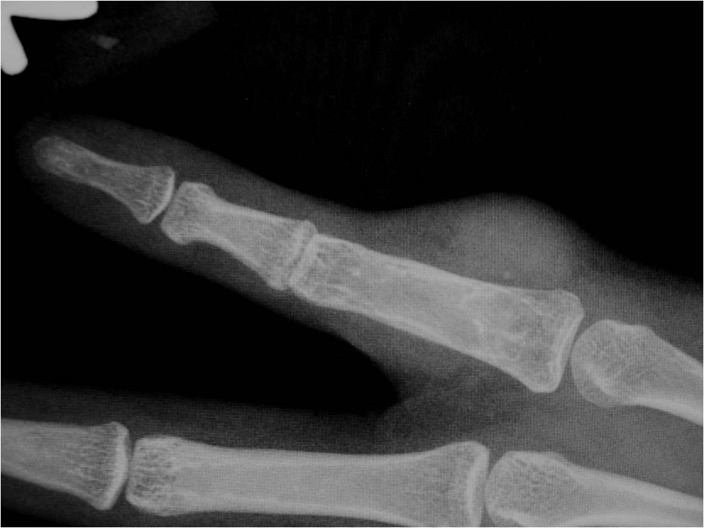 Fig. 1: Plain X ray of a hand with a giant cell tumor of tendon sheath demonstrates a soft tissue mass with no calcifications or bone infiltration. No pressure erosions of the cortical bone are seen in this patient.
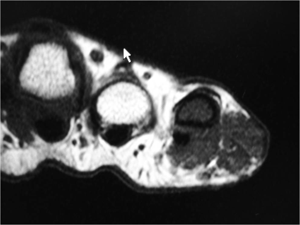
Fig. 2
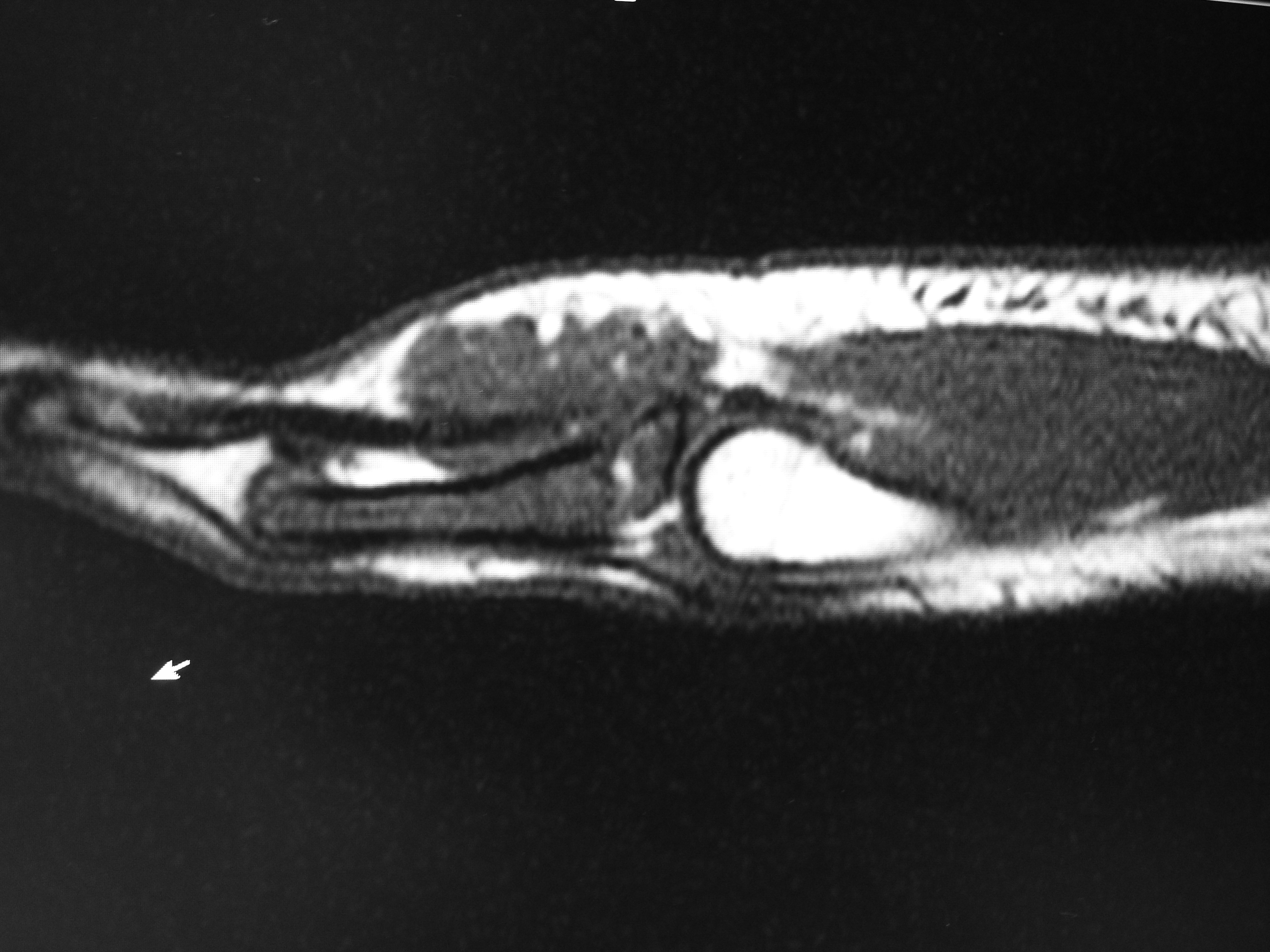
Fig. 3
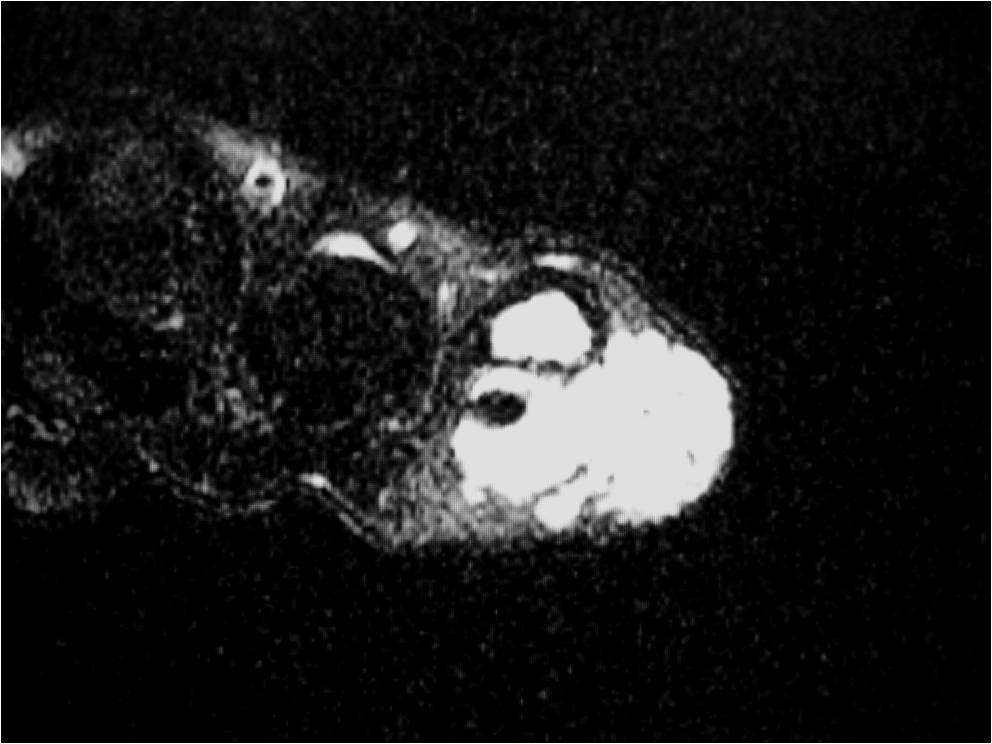
Fig. 4
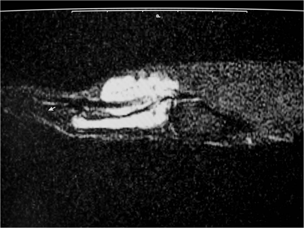
Fig. 5
Fig. 2-5: MR images of the hand with a giant cell tumor of tendon sheath shows a mass isointense to muscle on T1W and intermediate to high signal on T2W images. The lesion does not infiltrate the bone.
PATHOLOGY
Gross
•
Usually under 4 cm
• Can be larger in foot or near ankle
• Circumscribed
• May rarely erode bone and lose circumscription
• Often lobulated
• Cut section
• Usually white to gray
• Yellow and brown areas are common due to xanthoma cells and hemosiderin
Microscopic
•
Dense fibrous tissue divides tumor
• Gives nodular appearance
• Cells
• Small, round to oblong, often reniform or clefted nuclei
• Sometimes prominent nucleoli
• Oblong
• Frequently blend with spindled forms
• Variable numbers of giant cells
• Similar type of nuclei
• Usually contain 8-10 nuclei
• Xanthoma cells
• Sparse mitotic figures
• Low mitotic activity (3 to 5 mitosis x 10 HPF)
• Rarely necrosis is seen
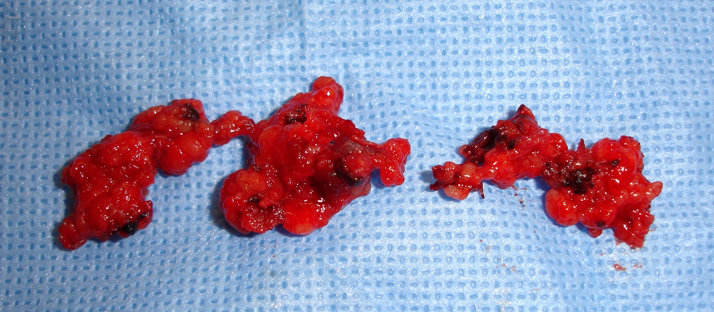
Fig. 6: Gross Pathology shows a lobulated and well circumscribed mass that is smaller than 5 cms.
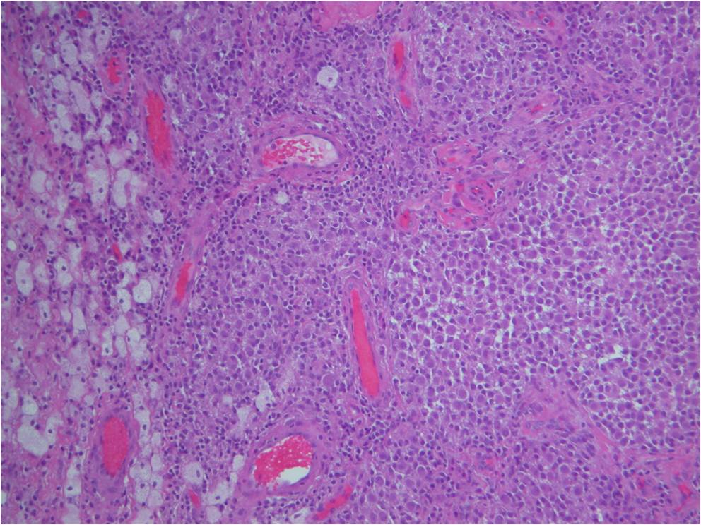
Fig. 7
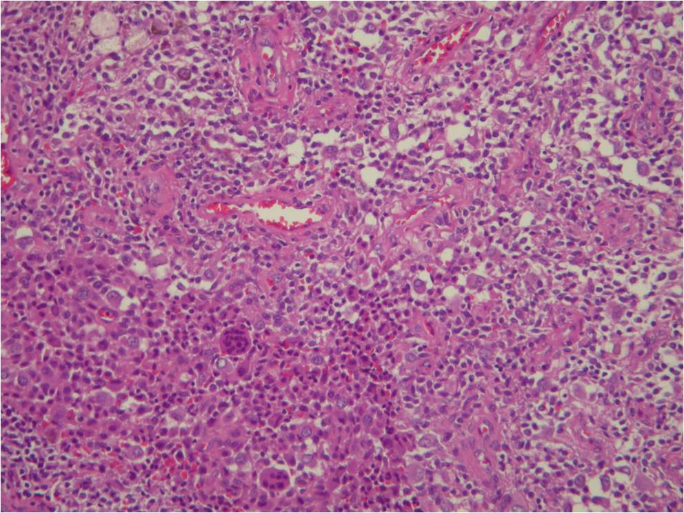
Fig. 8
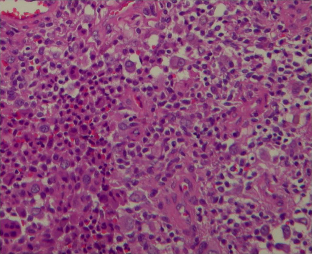
Fig. 9
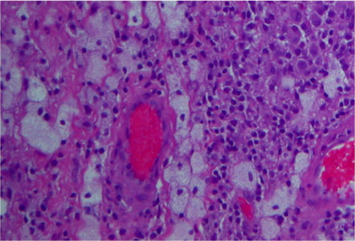
Fig. 10
Fig. 7-10: Microscopic pathology. Abundant small hystiocyte-like cells, numerous giant cells and xanthoma cells. In higher magnification images some foamy histiocytes are visible. No mitotic activity is present. Hemosidering deposition is common.
PROGNOSIS
Biological Behavior
• Rarely involve skin
• May erode bone infrequently
• Malignant change
• May occur (very rarely)
• Some reported cases
• Recurrence varies from 4% to 44%
• Recurrence rate is higher under some conditions, as;
• Diffuse, poorly encapsulated lesions
• Presence of satellite lesions
• Distal location in the digit
• Involvement of the extensor and flexor tendon and joint
• Intraosseous involvement
• Degenerative joint desease
• Rarely aggressive
• Does not metastasize
TREATMENT
• Complete surgical excision
• Recurrences can be reexcised
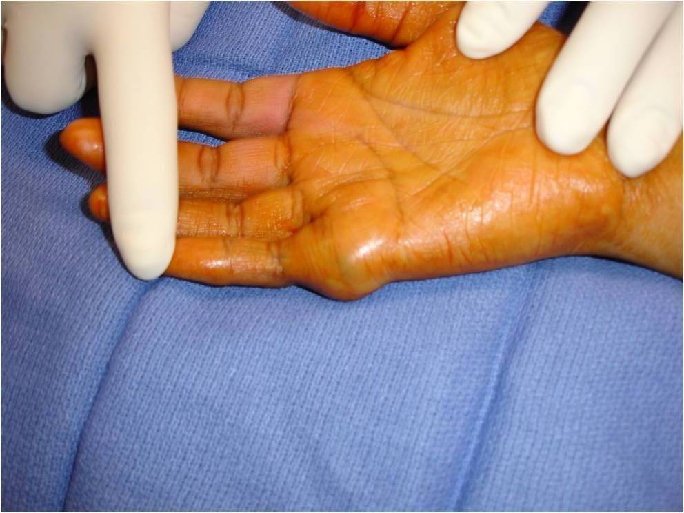 Fig. 11: Preoperative photo of the hand shows a large mass adjacent to the the 5th metacarpophalangeal joint area.
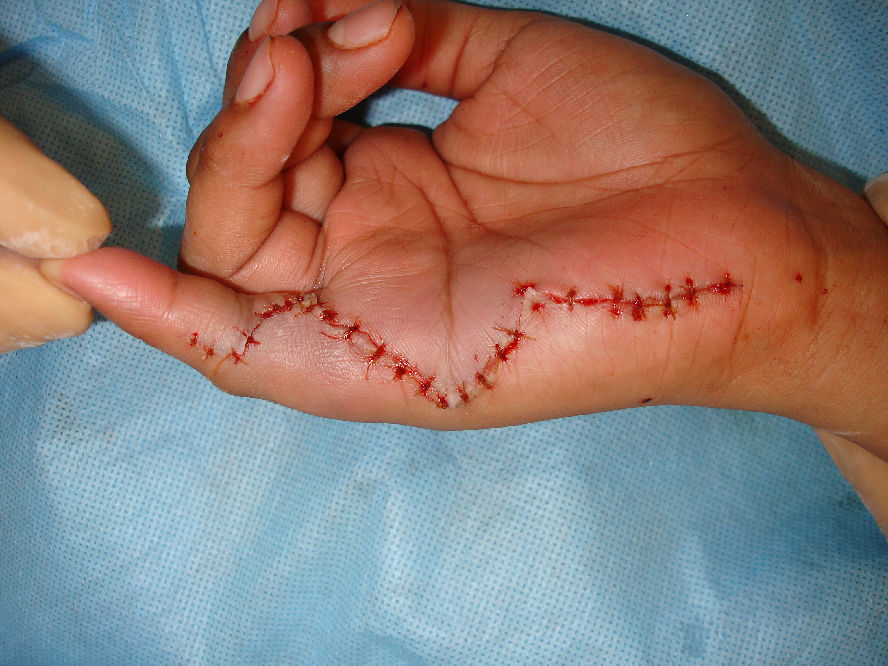
Fig. 12: Postoperative photo of the hand shows a sutured surgical incision in the hand after an extensive resection of a Giant cell tumor of the tendon sheath.
