GENERAL INFORMATION
Hibernoma is a benign neoplasm made up of multivacuolated brown fat cells. It is a type of lipoma.
CLINICAL DATA
•
Rare, represents less than 2% of all types of lipomas
•
Made of brown fat
•
Affects ages between second and third decade of life
•
Male preference
•
May produce steroid hormones
DIFFERENTIAL DIAGNOSIS
•
Lipoma
•
Liposarcoma (well-differentiated)
•
Rhabdomyosarcoma
•
Hematoma
CLINICAL PRESENTATION
Sign/Symptoms
•
Slow-growing mass
•
Painless
Prevalence
•
Preference for males (60%)
•
May occur in other age groups as well, but usually arises between 20 and 30 years of age
Site
•
Most commonly scapular and intrascapular region
•
Thigh, chest wall, back, axilla and groin
RADIOGRAPHIC PRESENTATION
Plain x-ray
• No specific radiological features
CT
• Well-defined lesion
• Tissue attenuation intermediate between fat and skeletal muscle
MRI
• Well-defined mass
• High signal intensity similar to fat on T1W and T2W (Fig. 1 and 2)
• T1W shows areas of low signal intensity between that of fat skeletal muscle
• On fat suppression sequences show incomplete fat suppression.
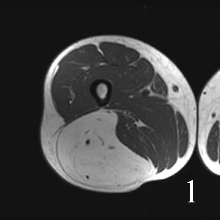
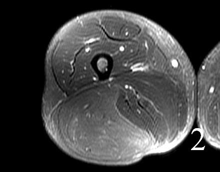
Fig. 1-2 MRI of lower extremity hibernoma (Fig. 1 and Fig. 2) shows similar signal intensity to subcutaneous fat fat on different sequences .
PATHOLOGY
Gross
•
Soft
•
Well-circumscribed
•
Tan to red-brown on cut section
•
Usually 5 to 10 cm Ø
Microscopic
•
Well-differentiated tumors, with lobulated pattern (Fig. 3)
Cells are uniform, round to oval cells
•
May have central nuclei
•
Granular to multivacuolated eosinophilic cytoplasm
Univacoulated fat cells are often admixed
•
Sometimes numerous enough to produce an appearance intermediate between hibernoma and ordinary lipoma
•
Poorly differentiated tumors
Endothelial cells are more atypical, close-packed, and often spindle-shaped
Progressive loss of evident vascular channels
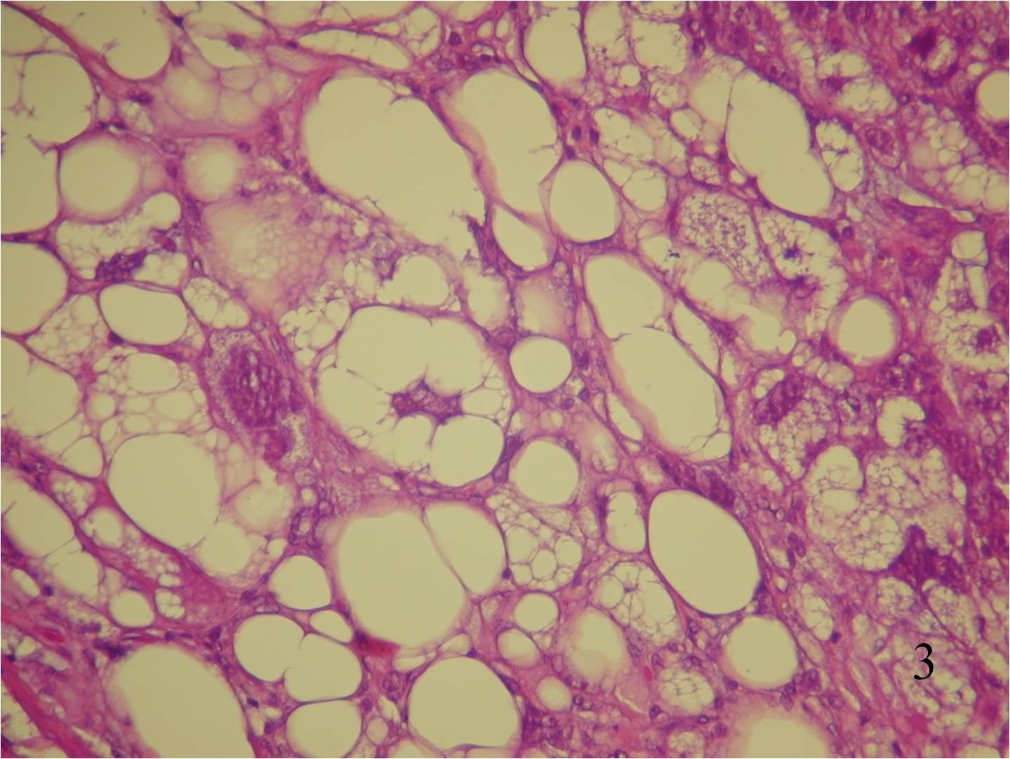 Fig. 3 High power photograph shows a well differentiated tumor, with uniform cells. Note the nuclei is small with no atypia. There are multivacuolated cells and a cell with a central nucleus
IMMUNOHISTOCHEMISTRY
Positive
•
S100
•
Oil red O
•
Sudan black
Negative
•
CD 34
•
P53
PROGNOSIS
BIOLOGICAL BEHAVIOR
•
Hibernoma is benign and does not metastasize or convert to a malignancy
•
It can grow to a large size
•
Non-invasive
•
Rare risk of recurrence after removal
TREATMENT
• Excision (Fig. 4 and 5)
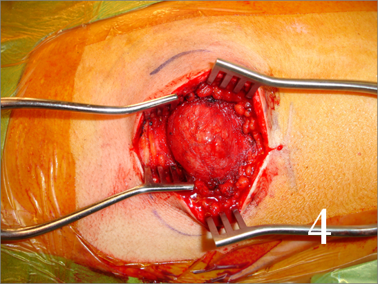
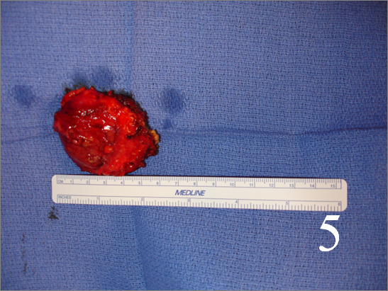 Fig. 4-5 Intraoperative photograph of a resection of a hibernoma (Fig. 4 and 5)
